|
OPS Imaging
Orthogonal Polarization Spectral
(OPS) Imaging represents a major innovation over conventional
intravital microscopy because of its portability and elimination of the
need for special preparations. Here's how it works:
Click here for animation: requires Flash
- Light from a source is
converted to a wavelength of 550 nanometers, the isobestic point for
hemoglobin. Hemoglobin becomes the contrast agent allowing for optimal
imaging of the microcirculation.
- The light passes through the first polarizer.
- The polarized light is directed towards the tissue by a set of lens.
- As the
light hits the tissue, it is reflected back through the lens. Most of
the light reflecting off the tissue and returning through the lens will
remain polarized. Ten percent or less of the light will penetrate
deeply into the tissue and go through multiple scattering events
becoming depolarized. The depolarized light is reflected back through
the lenses to a second polarizer or analyzer.
- The
analyzer, orthogonal (90 degrees) to the first polarizer, eliminates
the reflected polarized light and allows the depolarized light to pass
through to the CCD (Charged Couple Device), videocamera. The
depolarized light forms an image of the microcirculation on the CCD,
which can be captured through single frames or on videotape. The image
produced is as if the light source is actually placed behind the
desired target or transilluminated.
The Cytoscan® is the only
videomicroscope based on OPS imaging technology. Conventional
reflectance microscopy is of lower image quality, higher glare, and
reduced utility in clinical research settings. OPS Imaging has proven
to be equivalent to transillumination in terms of microscopy. Because
of its quality and portability, however the Cytoscan® allows researches
and clinicians to study microcirculation in humans in ways not possible
before.
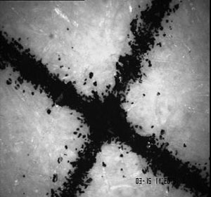
OPS-based image
|
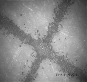
Conventional
|
|